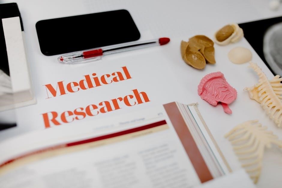
1.1 Overview of the Manual
This manual provides a comprehensive guide for human anatomy laboratory studies, focusing on cat dissections. It includes 30 exercises covering all body systems, with a clear and engaging writing style. Visual aids like summary tables and real-life examples enhance learning. The manual is designed for students in nursing, life sciences, and related fields, offering a hands-on approach to understanding human anatomy through comparative dissection.

1.2 Target Audience and Prerequisites
This manual is primarily designed for students enrolled in human anatomy and physiology courses, particularly those majoring in fields such as nursing, dental hygiene, occupational therapy, psychology, physical education, and life sciences. It is also suitable for students who require a foundational understanding of anatomy for their academic or professional pursuits. The exercises and activities are tailored to meet the needs of undergraduate students who may not have extensive prior knowledge of anatomy but are eager to gain practical, hands-on experience.
While no specific prerequisites are required to use this manual, students with a basic understanding of biological concepts, such as cellular structure and tissues, will find the content more accessible. Familiarity with scientific terminology and laboratory procedures is also beneficial but not essential, as the manual provides clear explanations and guidance. The manual assumes that students will have access to laboratory equipment and materials, including dissecting tools and specimens, as part of their course requirements.
The manual is structured to accommodate students with varying levels of preparation, offering visual guides, step-by-step instructions, and real-world applications to reinforce learning. It emphasizes the importance of safety protocols and proper laboratory techniques, ensuring that students can engage in dissection activities confidently and effectively. By the end of the course, students will have gained a comprehensive understanding of human anatomy through comparative dissection and hands-on exploration.

1.3 Key Features and Structure
The Human Anatomy Laboratory Manual with Cat Dissections by Elaine N. Marieb is meticulously organized to provide a comprehensive and engaging learning experience. The manual is divided into 30 exercises, each focusing on a specific aspect of human anatomy, from basic concepts to complex systems. Full-color illustrations and detailed diagrams are integrated throughout to help students visualize anatomical structures and their relationships. The inclusion of Visual Summary Tables simplifies complex information, making it easier for students to review and retain key concepts.
A unique feature of this manual is the “Why This Matters” boxes, which connect laboratory activities to real-life or clinical scenarios. This not only enhances understanding but also emphasizes the practical relevance of anatomical knowledge. The exercises are designed to be hands-on, with step-by-step instructions for dissections and other lab activities. Students are encouraged to engage actively with the material, fostering a deeper appreciation of human anatomy.
The manual’s structure aligns with most human anatomy and physiology courses, ensuring a logical progression from foundational topics to advanced studies. Each exercise builds on the previous one, creating a cohesive learning path. Additionally, the use of cat dissections provides a comparative approach, allowing students to explore anatomical similarities and differences between cats and humans. This comparative method enhances the learning experience and helps students develop a broader understanding of anatomical principles.
Overall, the manual is designed to cater to diverse learning styles, offering a combination of visual, textual, and practical elements. Its clear, engaging writing style and comprehensive coverage make it an indispensable resource for students seeking to master human anatomy through laboratory exploration.

2.1 Preparing for the Lab Environment
Preparing for the lab environment is crucial to ensure a safe, efficient, and productive learning experience. Before beginning any dissection or laboratory activity, students should familiarize themselves with the workspace and the tools required. The lab manual emphasizes the importance of organization and proper setup to avoid accidents and maintain a clean working area.
Key steps include donning appropriate personal protective equipment (PPE), such as gloves and lab coats, to minimize exposure to biological materials. Students should also ensure that all dissecting tools, such as scalpels, forceps, and dissecting scissors, are sharpened and within easy reach. The cat specimen should be properly thawed and positioned on the dissecting tray, with draping or paper towels used to maintain moisture and prevent contamination.
Understanding the lab layout is equally important. Students should locate essential resources, such as sinks, waste disposal bins, and emergency eyewash stations. Proper lighting and ventilation are critical to create a comfortable and safe working environment. The manual also recommends reviewing the lab schedule and objectives beforehand to stay focused during the session.
Finally, students are encouraged to review safety protocols and emergency procedures, such as handling sharp instruments and managing spills. By following these preparatory steps, students can create a conducive environment for active learning and exploration in the lab. Proper preparation not only enhances the quality of the lab experience but also ensures the well-being of everyone involved.
2.2 Safety Protocols and Emergency Procedures
Safety protocols are paramount in the anatomy laboratory to protect students and ensure a secure learning environment. The manual emphasizes adherence to specific guidelines to minimize risks associated with handling biological specimens and dissecting tools. Proper use of personal protective equipment (PPE), such as gloves and lab coats, is mandatory to prevent exposure to biological materials. Students are also advised to tie back long hair and avoid loose jewelry that could pose a hazard.
Handling sharp instruments, such as scalpels and dissecting knives, requires extreme caution. The manual instructs students to always cut away from their bodies and never pass sharp tools directly; instead, they should place them on a designated surface. In case of a cut or puncture wound, immediate action is required, including applying direct pressure, cleansing the wound, and reporting the incident to the instructor.
Emergency procedures are clearly outlined to address potential incidents. Eyewash stations and fire extinguishers must be easily accessible, and students should know their locations before starting lab work. Spills of preservatives or other chemicals should be contained and cleaned up immediately using appropriate materials. The manual also stresses the importance of proper disposal of biological waste and sharps to maintain a safe environment.
Regular safety drills and reviews of emergency protocols are recommended to ensure preparedness. Students are encouraged to report any unsafe conditions or equipment malfunctions promptly. By adhering to these safety protocols and understanding emergency procedures, students can contribute to a secure and efficient lab environment. Safety is everyone’s responsibility and should never be compromised.
2.3 Maintenance and Care of Lab Equipment
Proper maintenance and care of laboratory equipment are essential for ensuring longevity, functionality, and safety. This section outlines the steps and best practices for maintaining the tools and instruments used in human anatomy labs, particularly those involving cat dissections. Regular cleaning and disinfection of equipment, such as scalpels, forceps, and dissecting trays, are crucial to prevent contamination and bacterial growth. Students should use mild detergents and distilled water for cleaning, avoiding abrasive materials that could damage surfaces.
Sharpening and storing dissecting tools properly are vital. Dull instruments can cause accidents and reduce precision, so they should be sharpened periodically and stored in protective cases. Microscopes and other optical equipment require special care, such as covering lenses to prevent dust accumulation and calibrating adjustments regularly. Electrical equipment must be inspected for frayed cords or damaged plugs before use.
Lab surfaces, including dissecting tables and trays, should be wiped down with appropriate disinfectants after each use. Preserved specimens and biological materials must be stored in sealed containers to maintain their integrity and prevent odors. Proper labeling of all equipment and specimens is essential for organization and safety.
Students are encouraged to report any damaged or malfunctioning equipment to the instructor immediately. A maintenance schedule should be followed to ensure tools are serviced regularly. By adhering to these care and maintenance protocols, students can contribute to a well-organized, efficient, and safe laboratory environment. Proper upkeep not only extends the life of equipment but also ensures accurate and effective learning outcomes.

3.1 Basics of Anatomical Terminology
Mastering anatomical terminology is fundamental for understanding human anatomy and effectively communicating in scientific and medical contexts. This section introduces the basic terms and concepts used to describe the structure and organization of the human body. Anatomical terminology provides a universal language, ensuring clarity and precision in describing body structures, their locations, and relationships.
The foundation of anatomical terminology lies in understanding the anatomical position, the standard reference point for describing the body. In this position, the individual stands upright with feet shoulder-width apart, hands by the sides with palms facing forward, and eyes directed forward. This position allows for consistent and accurate descriptions of body regions and orientations.
Key directional terms are essential for locating structures within the body. These include anterior (front), posterior (back), dorsal (toward the back), and ventral (toward the abdomen). Additional terms like proximal (closer to the body’s center) and distal (farther from the center) help describe relative positions. Understanding these terms is critical for accurately identifying and discussing anatomical structures.
Body planes and regions are also central to anatomical description. The body can be divided into sagittal (front-to-back), frontal (side-to-side), and transverse (horizontal) planes, providing frameworks for visualization and dissection. Regions such as the cephalic (head), thoracic (chest), and abdominal (belly) areas further organize the body for study and communication.
Grasping these fundamental concepts ensures a strong foundation for exploring human anatomy, particularly in the context of cat dissections, where comparative anatomy is key. Clear understanding of anatomical terminology enhances both individual study and collaborative discussions, making it an essential skill for success in the laboratory and beyond.
3.2 Understanding Anatomical Position and Planes
Anatomical position and planes are fundamental concepts in human anatomy that provide a standardized framework for describing the body’s structure and orientation. The anatomical position serves as the reference point for all descriptive terminology. In this position, the body is upright, with feet shoulder-width apart, palms facing forward, and thumbs pointing toward the toes. This stance ensures consistency in describing body regions and directions.
The body can be divided into three main planes: sagittal, frontal, and transverse. The sagittal plane runs vertically from the top of the head to the bottom of the feet, dividing the body into left and right halves. The frontal (or coronal) plane also runs vertically but divides the body into front (anterior) and back (posterior) portions. The transverse plane is horizontal, cutting the body into top (superficial) and bottom (deep) sections.
Understanding these planes is crucial for visualizing and describing anatomical structures, especially during dissection. For example, a sagittal section of the brain reveals its internal structures, while a frontal section of the chest cavity exposes the heart and lungs. These planes also guide surgical procedures and medical imaging, such as MRI and CT scans.
Mastering anatomical position and planes enhances precision in communication and ensures clarity when describing complex anatomical relationships. This knowledge is particularly valuable in laboratory settings, where accurate terminology and spatial understanding are essential for successful dissection and analysis. These concepts form the cornerstone of anatomical study, enabling students to navigate the human body with confidence and accuracy.
3.3 Directional Terms and Body Regions
Directional terms and body regions are essential for precisely describing the location and orientation of anatomical structures. These terms provide a common language for communication in scientific and medical fields, ensuring clarity and accuracy. Common directional terms include anterior (front), posterior (back), superior (toward the head), inferior (toward the feet), medial (toward the midline), and lateral (away from the midline). Additionally, terms like proximal (closer to the origin) and distal (farther from the origin) describe locations along a structure.
The body is divided into distinct regions to simplify study and reference. The abdominal region covers the area between the ribs and pelvis, while the thoracic region encompasses the chest cavity. Other key regions include the cranial (skull), cervical (neck), lumbar (lower back), and pelvic areas. These regions help in localizing organs and structures, such as identifying the liver in the abdominal region or the heart in the thoracic region.
Understanding directional terms and body regions is vital for accurate dissection and identification of anatomical structures. For example, during a cat dissection, recognizing the ventral (belly) and dorsal (back) surfaces aids in locating internal organs. These concepts also apply to human anatomy, where they are used in clinical settings for diagnoses and treatments. By mastering this terminology, students can effectively communicate and navigate the complexities of anatomical study, whether in a laboratory or professional setting. This foundational knowledge is crucial for building a comprehensive understanding of human and comparative anatomy.
4.1 Nervous System Fundamentals
The nervous system is a complex control system that coordinates and regulates body functions. It consists of the Central Nervous System (CNS), which includes the brain and spinal cord, and the Peripheral Nervous System (PNS), comprising nerves that connect the CNS to the rest of the body. The CNS processes information and controls voluntary actions, while the PNS transmits sensory and motor signals.
The basic functional unit of the nervous system is the neuron, a specialized cell capable of transmitting nerve impulses. Neurons communicate through synapses, where chemical signals (neurotransmitters) are released and bind to receptors on adjacent neurons. This process enables rapid transmission of information throughout the body. Additionally, glial cells provide support and protection to neurons, ensuring their proper functioning.
The nervous system operates through reflexes, automatic responses to stimuli that do not require conscious thought. These reflexes are essential for immediate reactions, such as withdrawing a hand from a hot surface. The autonomic nervous system, a subdivision of the PNS, regulates involuntary functions like heart rate, digestion, and respiratory rate, operating through the sympathetic and parasympathetic divisions.
Understanding the fundamentals of the nervous system is crucial for studying anatomy, as it underpins the control and coordination of all bodily functions. This section provides a foundational overview, preparing students for more detailed explorations of nervous system structures and processes in subsequent chapters.
4.2 Muscular System and Movement
The muscular system is composed of specialized tissues that contract to produce movement, maintain posture, and regulate body temperature. It consists of three types of muscle tissue: skeletal, smooth, and cardiac. Skeletal muscles are attached to bones and are responsible for voluntary movements, such as walking or writing. Smooth muscles, found in the walls of internal organs like the digestive tract, function involuntarily, controlling processes like digestion. Cardiac muscle is exclusive to the heart and pumps blood throughout the body.
Muscles work in conjunction with the skeletal system to produce movement. Bones act as levers, while muscles and tendons function as the motive forces. The interaction between muscles and bones allows for a wide range of motions, from fine motor skills, such as picking up a pen, to gross motor activities, like running. Muscles also stabilize joints and maintain posture, preventing excessive movement that could lead to injury.
In the context of cat dissection, students observe the arrangement and function of major muscle groups, such as the flexors and extensors, which control limb movement. By examining the origins and insertions of muscles, students can understand how these tissues generate movement and maintain structural support. This hands-on approach reinforces the importance of the muscular system in enabling daily activities and maintaining overall bodily function.
Understanding the muscular system is essential for appreciating how the body moves and functions. This section provides a foundational exploration of muscle structure, function, and their role in movement, preparing students for further study of the musculoskeletal system.
4.3 Circulatory System and Blood Flow
The circulatory system, also known as the cardiovascular system, is responsible for transporting oxygen, nutrients, and hormones to cells and removing waste products. It consists of the heart, blood vessels, and blood. In the human body, the circulatory system is a closed system, where blood is confined within vessels and circulates throughout the body.
The heart is a muscular organ that pumps blood through the vessels; It has four chambers: the left and right atria, and the left and right ventricles. The right side of the heart receives deoxygenated blood from the body and pumps it to the lungs, while the left side receives oxygenated blood from the lungs and pumps it to the rest of the body. This separation ensures efficient oxygenation of blood.
Blood vessels include arteries, veins, and capillaries. Arteries carry oxygenated blood away from the heart, while veins return deoxygenated blood to the heart. Capillaries are the smallest vessels, where exchange of oxygen, nutrients, and waste products occurs between blood and tissues.
In the cat dissection, students observe the structure and organization of the circulatory system. By examining the heart and major blood vessels, students can understand the pathways of blood flow and the role of each component in maintaining proper circulation. This hands-on exploration complements theoretical knowledge, providing a deeper understanding of how the circulatory system functions in maintaining life and overall health.
Understanding the circulatory system is crucial for recognizing its role in overall health and disease prevention. This section provides a detailed examination of the system’s components and their functions, emphasizing the importance of blood flow in sustaining life.
4.4 Respiratory System and Gas Exchange
The respiratory system is essential for exchanging oxygen and carbon dioxide between the environment and the body. It includes the nose, pharynx, larynx, trachea, bronchi, and lungs. The primary function of this system is to facilitate gas exchange, which occurs in the alveoli of the lungs.
In the lungs, oxygen from inhaled air diffuses into the blood through the alveolar-capillary membrane, while carbon dioxide, a waste product, diffuses out of the blood and into the alveoli to be exhaled. This process is critical for cellular respiration and energy production.
In the cat dissection, students examine the respiratory organs, including the trachea, bronchi, and lungs, to understand their structure and function. The diaphragm, a major muscle involved in breathing, is also studied. Observing the alveoli and capillaries in the lungs provides insight into the microscopic structures responsible for gas exchange.
Understanding the mechanics of breathing and the pathways of air and blood is vital for comprehending how oxygen is delivered to tissues and how carbon dioxide is removed. This section emphasizes the importance of the respiratory system in maintaining homeostasis and overall health.
Through hands-on dissection and observation, students gain a deeper appreciation for the intricate design of the respiratory system and its role in sustaining life. This knowledge is foundational for understanding human anatomy and physiology, particularly for those pursuing careers in healthcare and life sciences.
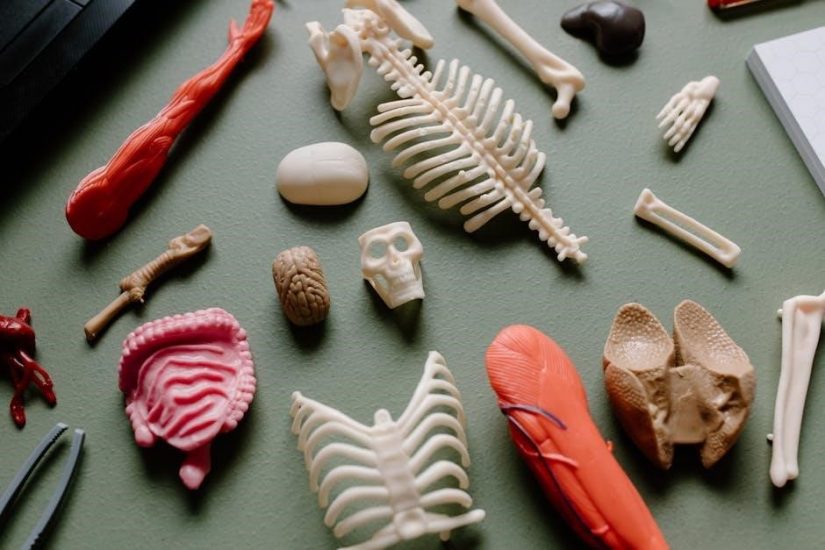
5.1 Preparing for Cat Dissection
Preparing for a cat dissection is a critical step to ensure a safe, effective, and respectful learning experience. Before beginning, students should thoroughly review the laboratory manual and understand the objectives of the dissection. Familiarizing oneself with the anatomical terminology and the tools required is essential.
The dissecting area should be clean and well-ventilated to prevent the spread of odors and maintain a comfortable working environment. Students should wear appropriate personal protective equipment (PPE), including gloves, lab coats, and eye protection, to minimize exposure to preservatives and other chemicals.
The cat specimen should be handled with care to avoid damage to its tissues. Proper positioning of the specimen on the dissecting tray is crucial for easy access to the organs and systems to be studied. Students should also ensure that all dissecting instruments, such as scalpels, forceps, and scissors, are sterilized and within reach.
Understanding the ethical considerations of dissection is equally important. Students should approach the activity with respect for the specimen and an appreciation for its role in advancing anatomical knowledge. Additionally, adherence to lab safety protocols and proper disposal of waste materials is mandatory.
Before starting the dissection, students should review the relevant chapters in the laboratory manual to gain a clear understanding of the cat’s anatomy. This preparation will help identify key structures and ensure a productive and educational experience.
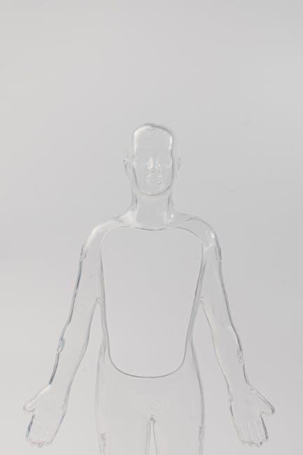
5.2 External Anatomy and Surface Features
The study of external anatomy and surface features is the first step in understanding the structural organization of the cat. This section focuses on identifying and describing the visible structures and their functions. Students begin by observing the cat’s overall body shape and noting the position of limbs, head, and tail. Key surface features, such as the skin, fur, eyes, ears, nose, and mouth, are examined in detail.
Students learn to identify specific anatomical landmarks, including the dorsal and ventral surfaces, as well as the lateral and medial aspects of the limbs. The manual emphasizes the importance of palpation to locate underlying structures, such as bones, muscles, and joints, without causing damage to the specimen. This hands-on approach helps students develop a deeper understanding of how external features relate to internal anatomy.
The section also covers the observation of natural behavior and reflexes, providing insights into the cat’s nervous system and sensory functions. Visual aids, such as detailed illustrations and photographs, are provided to assist in identifying complex structures. Comparisons with human anatomy are included to highlight evolutionary similarities and differences.
By thoroughly examining the external anatomy, students gain a solid foundation for the subsequent internal dissection. This exercise reinforces the importance of careful observation and proper technique in anatomical studies. The manual’s clear instructions and engaging visuals ensure a comprehensive understanding of the cat’s surface features and their significance in the broader context of anatomy.
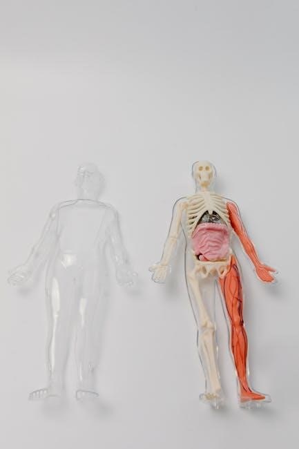
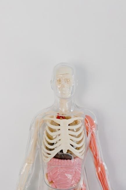
5.3 Internal Organ Dissection and Identification
This section guides students through the detailed dissection and identification of internal organs in the cat, providing a comprehensive understanding of their structure, function, and relationships. The manual begins with instructions on how to carefully open the abdominal and thoracic cavities, ensuring minimal damage to surrounding tissues. Students are then directed to locate and identify major organs, such as the heart, lungs, liver, stomach, intestines, kidneys, and reproductive organs.
Clear illustrations and photographs are provided to help students distinguish between organs and understand their spatial arrangement. The manual emphasizes the importance of recognizing anatomical landmarks, such as the diaphragm, ribcage, and pelvic cavity, to facilitate accurate dissection. Comparative notes between feline and human anatomy are included, highlighting similarities and differences in organ structure and function.
Students are also instructed on how to handle delicate tissues and use appropriate tools to avoid damaging organs during dissection. The section includes exercises on observing organ textures, colors, and vascular supply, which are critical for understanding physiological processes. Special attention is given to the digestive, circulatory, and respiratory systems, with detailed descriptions of their components and interactions.
Throughout this section, the manual encourages students to correlate their findings with previous studies of external anatomy and surface features. This integrative approach reinforces the connection between form and function, providing a holistic understanding of feline anatomy. The hands-on nature of this exercise prepares students for advanced studies in human anatomy and related fields.